BOTANIC LABORATORY MICROSCOPE IMAGES
- kubratas1582
- May 8, 2025
- 4 min read
Updated: Jul 10, 2025
First of all, the first material we will examine is ...''onion''... In general, this is what it looks like under the microscope
Let's take a closer look at this...

...................................................................................................................................................
The Latin name of the onion is ALLIUM CEPA, we usually encode materials with their Latin names because wherever we go in the world, the language of science is the same everywhere.
In terms of the way it is written, it has an epithet that first indicates the genus name and then the specific species. The genus name should be capitalized and the epithet should be lowercase, I think I was a little too excited in the early years of uni:)
Moving on, the examination is usually carried out by adding liquids, it depends on what you want from this experiment, but in general, we use pure water (I will talk about what pure water means later, now let's get back to our topic)
In microscope imaging, the magnification coefficient is indicated by expressions multiplied by ten
For example, here it is written as four times ten... this.... It means that the microscope shows the material at forty times magnification, and when asked how many times we have examined it, we say that we have examined it in "fourth". Likewise, we examine the tenth, fortieth, and hundredth, and in the hundredth lens, a magnification of a thousand times occurs. The reason is that microscopes have two lenses ... We call the lens in the upper part close to our eye the ocular lens... The material we use, that is, the part close to the object, is called the objective lens. The ocular lens always magnifies the object tenfold, while the objective lens is in the changing part. These magnifications may vary according to the microscopes used, but if we want to explain them in terms of the way they are written and the way they are pronounced, this is the basic logic.
Below is a schematic drawing of a plant cell, and a plant cell has a cell wall, unlike animal cells. When we look a little inside the cell, we come across a middle lamella.
The middle lamella are normally located outside the cell wall in plant cells. A phenomenon that occurs during cytoplasm division and is subsequently involved in the formation of the wall of the new cell.
Its main component is pectin, which ensures the cohesion of cells and the durability of tissues.
Providing intercellular mechanical support and maintaining the integrity of plant tissues is the main task of the middle lamella.
Then, after passing the inner cell membrane, we encounter organelles and a nucleus. We use Lugol's solution to stain these cell and tissue samples. This solution allows the cell nuclei and structures within the cell to be seen more clearly.
What supports the entry of Lugol into the cell is the "VACUOLE" organelle.
Plant cells usually have a large central vacuole. This vacuole maintains the water balance in the cell, regulates cell turgor (pressure) and stores waste materials.
If we take a closer look. The vacuole stores most of the water taken into the cell by passing through the passages that provide the transport of intercellular substances and provides support to the cell wall thanks to the pressure of the water on the plant cell. This pressure allows the plant to stand upright and we call this pressure Turgor Pressure.

...................................................................................................................................................
Continuing, this is a closer view of the microscope image obtained from the previous onion skin… the cell nucleus and cell wall are more clearly visible thanks to the Lugol solution

...................................................................................................................................................
After each viewing, we draw what we see to make it more memorable. This is my first drawing experience :D

...................................................................................................................................................
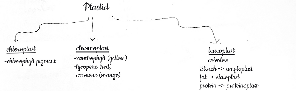
YES, it's a new topic... plastids,
Plastids are double-membrane-enclosed organelles found in plant cells and some algae and take on many vital functions. Plastids play an important role in processes such as photosynthesis, energy storage and "pigment production". There are various types of plastids that are specialized according to different functions, and these plastids can transform into each other during the development of the plant.
If we look at the Plastid Types, they are divided into three
First, let's take a look at the Chloroplast:
Chloroplasts are plastids that carry out photosynthesis in plant cells. They contain chlorophyll pigment, which absorbs sunlight and allows the plant to produce nutrients.
Inside the chloroplasts are thylakoid membrane systems, which are a special internal structure involved in photosynthesis. This structure is responsible for the production of ATP and provides energy for the plant.
Chromoplasts as a second type of plastid:
..., are plastids that are found in parts of plants such as flowers, fruits, leaves and contain red, orange and yellow colors in these regions.
These plastids contain pigments such as xanthophyll, lycopene and carotene. Xanthophyll is yellow, lycopene is red, and carotene is orange.
and they create bright colors to encourage pollination or seed dispersal of plants.
The last type of plastid, the... Leucoplasts :
They are colorless plastids and are found in tissues that do not receive light, such as roots and underground stems in plants.
First tyoe of leucoplast is Amyloplast: It is a type of leucoplast that stores starch.
Second type of leucoplast is Elaioplast: It is a type of leucoplast that stores olil.
And last type of leucoplast is Proteinoplast: It is a type of leucoplast that stores protein.
...................................................................................................................................................
Plastid, which we will examine first, is a chromplastic containing lycopene pigment obtained by scraping method from tomato fruit, whose Latin name is Lycopersicum Esculantum.

...................................................................................................................................................
This is what it looks like on the ten times magnification

...................................................................................................................................................
In fourty times magnification, it is like this

...................................................................................................................................................
And this is my drawing

...................................................................................................................................................
Our second material: Carrots ... Its latin name is Daucus carota ... We took a cross-section from the circular part and when we examined it under the microscope, we got an image like this :

...................................................................................................................................................
The name of our chromplast here is carotene .... It does not appear very clearly, since it is concentrated in the right part, but if we look at the central parts, we can see small, elongated and scattered pigments ... Here's one of them.

...................................................................................................................................................
This is another visual, in fact, it is the same as before, the only difference is the angles of receiving light
...................................................................................................................................................
Now let's move on to the other material
Our next plant is ... telegraph flower .... Its Latin name is Tradescantia sp.
Sectioning Shape: Underleaf Superficial... Here we aim to see a leukoplast, …let's look at our microscope images at three different proximities to observe our plastids, which is a type of plastid with a transparent structure.

...................................................................................................................................................
The version in this 4x ... Here you can see a lot of leukoplasts ...

...................................................................................................................................................
And this is the case with the 10x...

...................................................................................................................................................
And this is what it looks like when you zoom in even closer.

...................................................................................................................................................
And our final plastid review material is a green algae.... Its Latin name, Klodopora sp. .... There is no sectional method:)
And what we're hoping to see here is chlorophylls that contain the pigment chlorophyll

...................................................................................................................................................
that's what it looks like... how intense isn't it....

...................................................................................................................................................
This is what it looks like a little closer...

...................................................................................................................................................
The next things we're looking at are kind of lifeless stuff..... I think the name is on it, we call these structures in the cell ergastic substances. and Ergastic substances, which we will examine.... Starch grains...
Starch grains are divided into two .... According to the location of the hilum, it is called centric and ectric. and according to the morphological structure and arrangement of starch grains, it is divided into three. They can be observed as simple, compounds and semi-compounds.
the first material we examined ... potato... Its Latin name is Solanum tuberasum... We examined the sections obtained by the scraping method in tens and forty lenses

...................................................................................................................................................
If we look at this image, we can call it a "simple centric" starch grain because the starch grain in the middle is located in the center of the hilum - umbilicus, or in other words, the eye, and has a simple structure.
If we look at the grains right next to it or on the upper right side, they are called "simple ectric" starch grains.
If we look at the top left, we can see a "semi-compound ectric" starch grain here

...................................................................................................................................................
I wanted to take a closer shot of the semi-compound ectric starch grain... Known... It's a hard thing to find :)

...................................................................................................................................................
Our other material is ... bean... Its Latin name is Fascinius vulgaris...
we expected this material to look like under the microscope

...................................................................................................................................................
This is how the sections taken by the scraping method appear in the forty times magnification
If we turn the brightness down a bit, in

...................................................................................................................................................
this way we can get a clearer image. Here are simple grains of centric starch in beans ...

...................................................................................................................................................
Our second material was corn and the morphological structure we wanted to see was supposed to look like this, but we couldn't grow it because it was a very difficult material to scrape, or I don't remember:) So let's get past that:d

...................................................................................................................................................
Our last material is rice . Its Latin name is Oryza Sativa... When we examine it in forty times magnification with the scraping method, we aim to see simple or compound centric starch grains.

...................................................................................................................................................
Yes, this was the clearest image we were able to capture under the microscope, and in the center you can see the grains of compound centric starch. Thank you for listening to me. see you later.

References:
Gallei, M., Truckenbrodt, S., et al. (2024). How expansion microscopy can be applied to plants. Plant Cell, 37(2), koaf002. https://doi.org/10.1093/plcell/koaf002
Komis, G., Šamajová, O., Ovečka, M., & Šamaj, J. (2015). Super-resolution microscopy in plant cell biology. Journal of Microscopy, 259(1), 1–9. https://doi.org/10.1111/jmi.12140
Lux, A., Benfey, P., & Beemster, G. T. S. (2005). Rapid preparation and examination of plant sections with microscopy. Motic Microscopes.
Roszak, P., Thomas, H., & Davies, J. (2019). Putting plants under the microscope. University of Cambridge.
Gage, S. H. (1904). The Microscope; An Introduction to Microscopic Methods and to Histology. Comstock Publishing Co.
Komis, G., Šamajová, O., & Šamaj, J. (2015). Imaging the living plant cell: From probes to quantification. Plant Methods, 11(1), 1–12. https://doi.org/10.1186/s13007-015-0071-8
Shaw, P. J. (2001). Introduction to optical microscopy for plant cell biology. In Plant Cell Biology, A Practical Approach (pp. 3–20). Oxford University Press.
https://link.springer.com/chapter/10.1007/978-0-387-45524-2_44


Comments